
Table of Contents
What is Four-Dimensional Ultrasound?
Four-dimensional ultrasound Turkey has become a popular choice among expectant parents who want a more detailed and emotional connection with their unborn baby. Unlike traditional methods, this advanced imaging technology captures real-time movements and facial expressions, allowing families to experience precious moments before birth. Thanks to its modern healthcare facilities and experienced professionals, Turkey is a top destination for families seeking four-dimensional ultrasound Turkey for both medical reassurance and bonding.
When is Four-Dimensional Ultrasound Done?
Four-dimensional ultrasound is usually done between 22 and 32 weeks of pregnancy. This period is a time when important organs and structures in the development of the fetus become evident and mobility increases. Four-dimensional ultrasound performed during this period provides the opportunity to examine the anatomical and physiological characteristics of the baby in detail. However, four-dimensional ultrasound can be performed outside this time period, in line with the doctor’s recommendation and certain medical requirements.
Four Dimensional Ultrasound Usage Areas
The usage areas of four-dimensional ultrasound are quite wide. Its most well-known use is fetal monitoring during pregnancy. Examinations performed with this technology help determine the baby’s development, gender, potential health problems and even some genetic problems. In addition, four-dimensional ultrasound provides the opportunity to record the facial expressions and movements of the baby in the womb as unforgettable moments for families, which helps strengthen the emotional bond between parents and the baby.
In terms of other medical applications, four-dimensional ultrasound allows detailed examination of intra-abdominal organs, especially organs such as the liver, kidney and gallbladder. In this way, structural anomalies of organs, cysts, tumors and other pathological changes can be observed more clearly.
Four-dimensional ultrasound, as one of the indispensable tools of modern medicine, provides healthcare professionals and patients with valuable information in the diagnosis and management of diseases. Thanks to this technology, medical examinations can be performed more effectively and efficiently, and at the same time the quality of patient care can be improved.
How Does 4D Ultrasound Differ from 2D and 3D?
Understanding the difference between 2D, 3D, and 4D ultrasound is essential for expectant parents making informed choices:
2D Ultrasound: This is the standard black-and-white imaging used to monitor fetal growth, heartbeat, and general development. It provides flat, cross-sectional views.
3D Ultrasound: Offers still, three-dimensional images of the baby, capturing features in greater detail, such as the face, hands, or feet.
4D Ultrasound: Adds the element of time to 3D images, displaying real-time video of fetal movements, including yawns, smiles, or stretching limbs.
The four-dimensional ultrasound Turkey experience is not just medical—it’s emotional, giving parents a first glimpse of their baby’s personality and behavior in the womb.
Why Do Expectant Parents Choose 4D Ultrasound?
Expectant parents choose four-dimensional ultrasound in Turkey for several personal and clinical reasons:
Emotional Bonding: Seeing your baby move, smile, or yawn enhances emotional connection between parents and their unborn child.
Family Sharing: These detailed images and videos can be shared with loved ones or kept as treasured keepsakes.
Medical Insight: In some cases, 4D imaging can help detect anomalies not visible in 2D scans, assisting with early intervention if needed.
Peace of Mind: High-resolution imaging provides reassurance about the baby’s development and well-being.
Memorable Experience: Many clinics in Turkey offer luxurious scanning environments, making the procedure a memorable and comforting experience.
Benefits of Having 4D Ultrasound in Turkey
Choosing four-dimensional ultrasound Turkey comes with multiple advantages:
State-of-the-Art Equipment: Clinics in Turkey are equipped with the latest ultrasound technology for the clearest, most detailed imaging.
Experienced Technicians: Skilled radiologists and sonographers ensure accurate scans and a comfortable process.
Affordable Pricing: Turkey offers significantly lower prices compared to many European and Western countries, without compromising on quality.
Hospitality and Comfort: Clinics often provide private rooms, calming environments, and family-friendly settings.
Quick Appointments: Short waiting times make it easy for local and international patients to schedule their ultrasound at a convenient time.
How is Four-Dimensional Ultrasound Done?
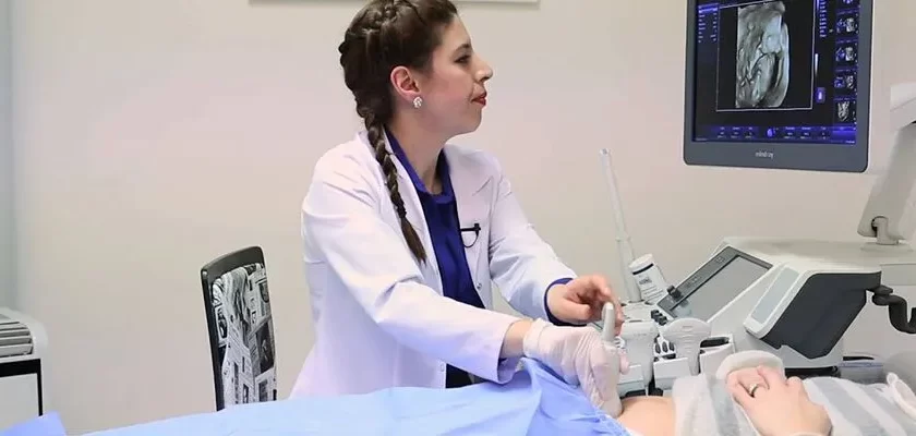
Is performed using specially designed ultrasound devices. These devices contain sensors and software with advanced imaging capabilities. During the procedure, the ultrasound probe is placed on the relevant area of the patient’s body, usually the abdomen. This probe emits high-frequency sound waves, and these waves are reflected in different ways by body tissues. The reflected sound waves are received again by the probe and processed by computer software to create three-dimensional images. Because these images are updated several times per second, they provide viewers with a vibrant, moving visual. For example, if the baby in the womb makes sucking movements, these movements can be followed moment by moment and observed in detail.
To whom is Four-Dimensional Ultrasound performed?
Four-dimensional ultrasound is generally the preferred method during pregnancy. It is used especially during the second and third trimesters of pregnancy to monitor the baby’s development in detail and to detect possible health problems early. In addition, it is also preferred to determine the facial expressions, movements and sometimes gender of babies. However, it is important to note that this technology is not necessary or appropriate for every pregnancy situation. The use of four-dimensional ultrasound depends on factors such as the health status of the mother and the fetus, gestational age, previous ultrasound results, and the physician’s recommendations . Additionally, in some cases, it can be done only at the request of the prospective parents, without medical necessity.
Are Four-Dimensional Ultrasound and Detailed Ultrasound the Same Thing?
The main difference between four-dimensional ultrasound and detailed ultrasound is in visual presentation and intended use. Detailed ultrasound is a procedure that is usually performed between 18 and 22 weeks of pregnancy and examines the fetus’s organs, skeletal structure and body contours in detail. This examination is critical to detect possible congenital anomalies or other health problems in the fetus. Four-dimensional ultrasound takes these detailed examinations one step further, adding a time dimension and showing the fetus’ movements and behaviors live. This provides a richer visual experience and provides an opportunity for parents to establish an emotional connection.
Four-dimensional ultrasound is one of the most advanced imaging technologies offered by modern medicine and provides valuable information for both parents and healthcare professionals, especially during pregnancy. However, it is important to remember that the use of this technology should always be done under physician supervision and based on appropriate medical recommendations.
What You Need to Know About Four-Dimensional Ultrasound
Four-dimensional ultrasound goes one step beyond three-dimensional ultrasound technology, providing images that move in real time. Thanks to this technology, the movements, facial expressions and sometimes even the sounds of babies in the womb can be observed. This method especially helps understand the development of babies, detects potential health problems early, and allows parents to bond with their babies earlier.
One of the biggest advantages of four-dimensional ultrasound is that it provides three-dimensional images of the fetus’s organs and structure in real time. This allows doctors to have more detailed information about the health status of the fetus. However, it is important to note that this type of ultrasound is not necessary or mandatory in every pregnancy. This technology is often used within recommended medical protocols and in certain situations.
Four-dimensional ultrasound may be more expensive than other standard ultrasound techniques and may not be available in all health centers. Therefore, before planning this procedure, it is important to obtain information about your means and health insurance coverage.
Preparation Process Before Four-Dimensional Ultrasound
There are several important points to consider when preparing for four-dimensional ultrasound. First, a preliminary consultation with your doctor is recommended before the ultrasound scan. This meeting focuses on topics such as the purpose of ultrasound, how it is performed, and what kind of information it can provide.
Four-dimensional ultrasound is usually done in the second trimester of pregnancy (usually between 24 and 32 weeks). This period is ideal for the fetus to be seen more clearly and its movements to be followed more clearly. On the day of an ultrasound, drinking plenty of water is often recommended because a full bladder can improve image quality by creating a better window for the ultrasound waves of the uterus.
You may be asked not to eat before the ultrasound, usually for a period of 8-12 hours, especially if the ultrasound procedure is scheduled for the morning. However, this may vary depending on the type of ultrasound to be performed and the doctor’s recommendations. Additionally, it is important that you wear comfortable clothing and assume a comfortable position during the procedure.
Finally, having all your medical records and previous ultrasound results ready before the four-dimensional ultrasound will help your doctor make a more comprehensive evaluation. This information can be used to better evaluate any changes or improvements by comparison with previous ultrasound results.
Difference of Four-Dimensional Ultrasound from Classical Ultrasound
Four-dimensional ultrasound provides moving images by essentially adding the “time” dimension to three-dimensional ultrasound images. This provides the possibility of observing instantaneous movements and dynamic changes, unlike classical two-dimensional or three-dimensional ultrasounds. Classical two-dimensional ultrasound usually displays tissues and internal organs as plane sections. Three-dimensional ultrasound creates a volume based on these sections, but these images are static and do not move. Four-dimensional ultrasound, on the other hand, gives the opportunity to live monitor instantaneous behaviors, such as a baby smiling or yawning in the womb.
These dynamic images provided by four-dimensional ultrasound allow doctors to make a more comprehensive evaluation. Particularly in prenatal diagnosis and monitoring of fetal health, four-dimensional ultrasound offers the opportunity to evaluate the baby’s movements, position changes and, in some cases, even the function of organs in real time.
Why is Four-Dimensional Ultrasound Desirable?
Four-dimensional ultrasound may be ordered for a variety of medical needs, especially for pregnancy monitoring. One of the most important areas of use of this technology is prenatal diagnosis. Four-dimensional ultrasound is preferred to monitor the baby’s development and health status in detail, detect potential health problems early and plan intervention when necessary.
This method is also extremely valuable, especially for parents who want to observe the baby’s movements and behaviors. Since babies’ facial expressions, movements and sometimes reactions can be seen clearly, emotional satisfaction and bonding process is supported for expectant parents. Four-dimensional ultrasound can also provide important information regarding the diagnosis of some structural abnormalities, such as cleft lip or spinal disorders.
On the other hand, the use of four-dimensional ultrasound may not always be a medical necessity. In some cases, this method can be used optionally for the sole purpose of satisfying parents’ curiosity or early bonding with their baby. However, as with any medical procedure, the use of four-dimensional ultrasound should be performed with the advice and guidance of a physician.
In conclusion, four-dimensional ultrasound stands out among advanced imaging techniques with its capacity to provide particularly detailed and dynamic information. This technology, which is a valuable resource for both doctors and parents, plays an important role in prenatal care processes by maximizing the opportunities offered by modern medicine.
Can We See the Baby in Four-Dimensional Ultrasound?
Four-dimensional ultrasound is a version of three-dimensional ultrasound with the added time dimension. This means that the images obtained during the ultrasound are moving and parents can see their baby moving in real time. Thanks to four-dimensional ultrasound, babies’ facial expressions, movements and sometimes position changes can be clearly observed. These detailed images, which show babies’ natural reflexes such as thumb sucking, yawning and smiling, can create very emotional and unforgettable moments for parents.
This technology also offers doctors the opportunity to better monitor the development of babies. Especially when it comes to developmental disorders or potential health problems, thanks to four-dimensional ultrasounds, doctors can diagnose problems early and prepare appropriate intervention plans more effectively.
Four-Dimensional Ultrasound Turkey Prices 2026
The cost of four-dimensional ultrasound Turkey in 2026 varies based on the clinic’s location, the length of the session, and any extras such as photos, DVDs, or printed reports. While pricing in Turkey remains far more competitive than in Western countries, quality should never be sacrificed for cost.
Each clinic may offer packages that include:
Basic imaging
Full video recording
Gender determination
Printed or digital photos
For an accurate quote and details tailored to your preferences, four-dimensional ultrasound Turkey prices 2026 – contact us now.